Electron microscopy provides new view of tiny virus with therapeutic potential
Phys.org | September 07, 2018
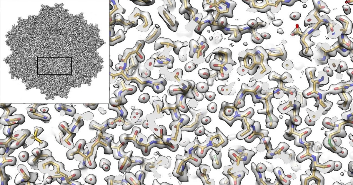
The imaging method called cryo-electron microscopy (cryo-EM) allows researchers to visualize the shapes of biological molecules with an unprecedented level of detail. Now, a team led by researchers from the Salk Institute and the University of Florida is reporting how they used cryo-EM to show the structure of a version of a virus called an AAV2, advancing the technique's capabilities and the virus' potential as a delivery vehicle for gene therapies.
"It's not an overstatement to say that this is one of the best cryo-EM structures that's ever been achieved in this field," says Salk Assistant Professor Dmitry Lyumkis, a structural biologist and co-senior author of the study. "We applied a number of different procedures that have previously only been described in theory. We demonstrated experimentally, for the first time, that they can be used to dramatically improve the quality of this kind of imaging."